Very frequent in women during reproductive period (25-45 years)
Can mimic several situations, clinically and radiologically
Usually multifocal and bilateral
Can present with discomfort, pain, heaviness or nipple discharge
Some cases can even present with axillary lymphadenopathy
Ultrasound usually discovers fibrosis, cysts and microcalcifications
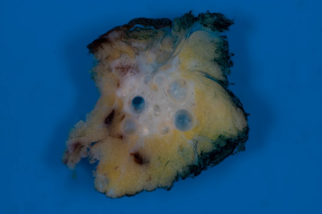
The gross appearence is a poorly defined, gray-white area with cysts of variable size
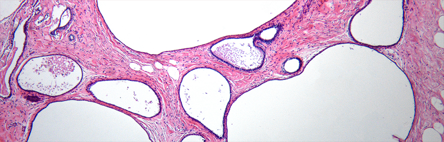
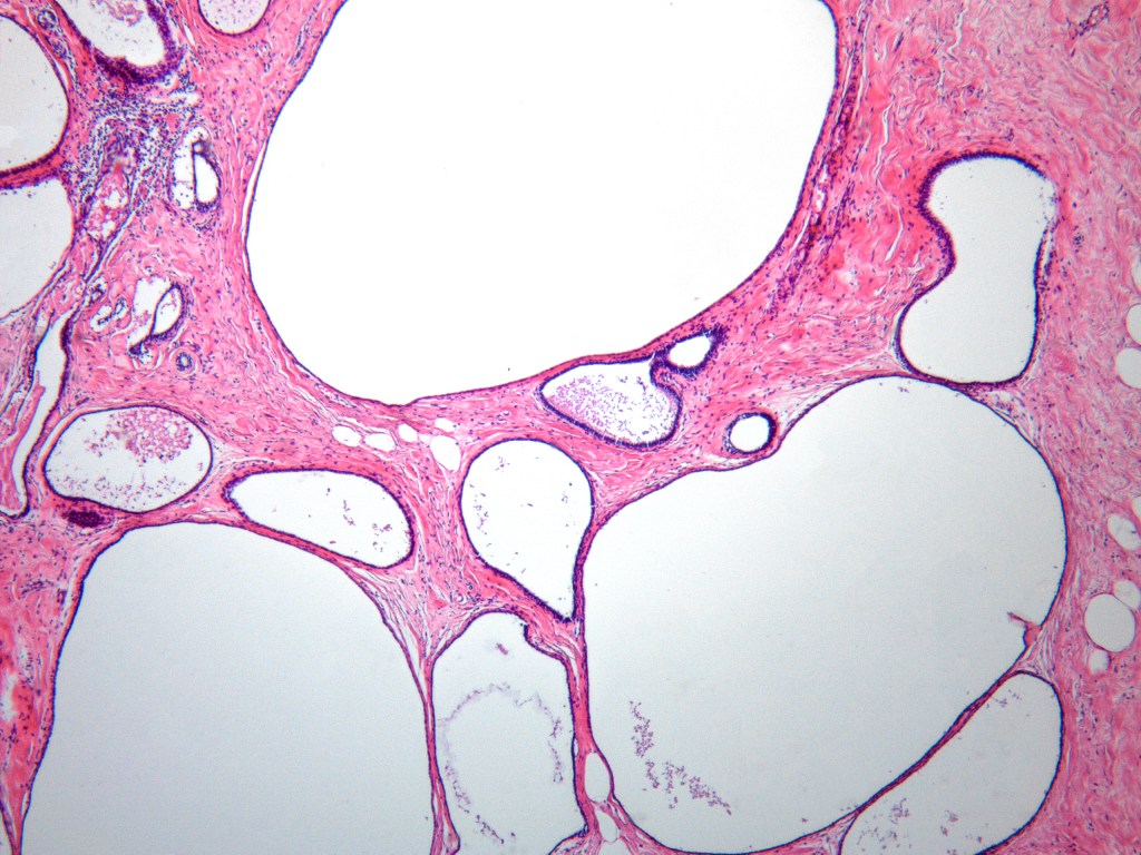
Microscopic presentation consists in a series of findings, some of them are:
- Cysts (cystic dilatation of ducts and acini), filled with eosinophilic material, foamy cytoplasm macrophages, blood, hemosiderin and sometimes microcalcifications
- Apocrine metaplasia (I’m thinking that cysts and apocrine metaplasia are the typical findings)
- Fibrosis (of course)
- Chronic inflammation
- Fibroadenomatoid changes
- Epithelial hyperplasia (if prominent should be called proliferative fibrocystic changes, denotating the associated risk)

Deja un comentario