Described in 1979 (by Enzinger), relatively new I would say, it usually affects children and young adults. Usual sites are extremities and trunk. The tumor usually locates in the deep dermis or subcutaneous tissue. Clinically, it can appear like a hematoma or vascular lesion. Sometimes, patients can experience fever or mild pain.
This tumor has low malignant potential, 15% can recur, and a minor 5% can metastasize.
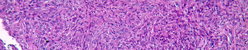
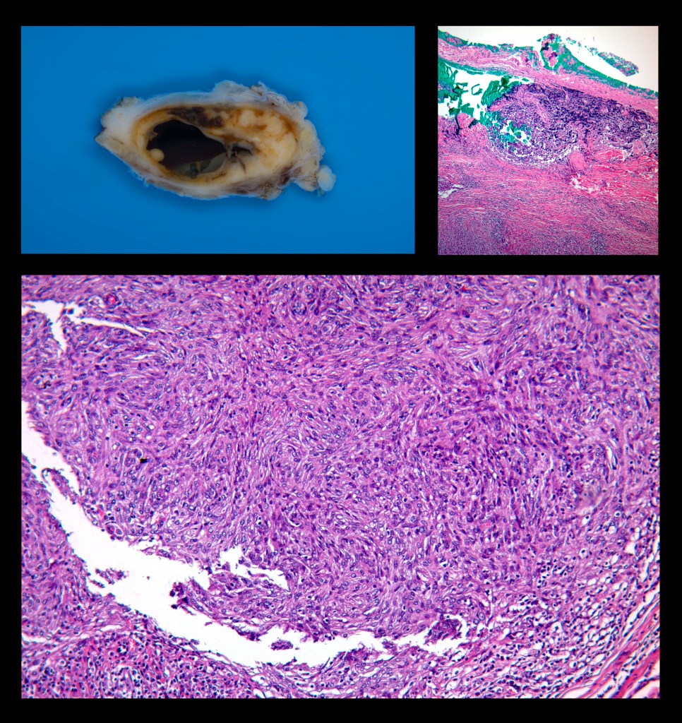
The gross appearance is a well-circumscribed tumor, solid-cystic, yellow to brown. Microscopically, the constant finding is ovoid to fusiform cells reminiscent of histiocytes. The typical cases show a pseudocapsule with prominent lymphocytic infiltrate, so much so that it can even simulate a metastatic lymph node, pseudovascular spaces and slit-like spaces, hemosiderin, giant cells, low atypia, and few mitoses.
This tumor presents rearrangements of the EWSR1 gene. Up to 90% of the cases show this particular fusion [EWSR1-CREB1 t(2:22)]; importantly, this fusion is not specific to this tumor. Other tumors with different morphology, presentation, and prognosis can share this fusion, which is something to keep in mind; molecular specificity is not guaranteed. This tumor is rare, so to me, this is the importance of sharing this information.

Deja un comentario